Equi-note 21 Hyperflexion-rollkur can damage the horse's neck causing osteoarthritis
Source: Library.academialberti.de
Equi-note 21
Hyperflexion-rollkur can damage the horse’s neck causing osteoarthritis
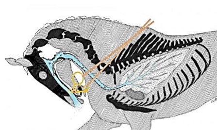
Previous posts have referred to damage created by constricting the trachea. This affects the horse’s ability to breathe and has an effect on the speed of the airflow. However, in addition, there are further areas where damage can occur.
This post refers to damage of a more structural nature, which occurs to the cervical joints themselves. While jumping and dressage are very different sports, considerable overlap exists in the training methods. Especially when it comes to rein length and rein pressure.
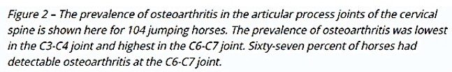
The following image shows where one researcher identified the location of cervical osteoarthritis in jumping horses.
Prevalence of Cervical Osteoarthritis in Jumping Horses
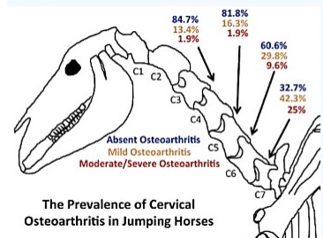
Source: Espinosa-Mur P, Phillips KL 1, Galuppo LD 1, DeRouen A, Benoit P, Anderson E, Shaw K, Puchalski S, Peters D, Kass PH 1, Spriet M 1. Radiological prevalence of osteoarthritis of the cervical region in 104 performing Warmblood jumpers. Equine Vet J. 2021 Sep;53(5):972-978. doi: 10.1111/evj.13383. Epub 2021 Jan 11. PMID: 33174228; 1-School of Veterinary Medicine, University of California, Davis, California, USA.
“Background: Cervical osteoarthritis (OA) has been documented as a potential source of pain and poor performance in sport horses.
Conclusions: This study showed that, although there is a range of interpretation of radiographic findings of the APJ, OA of the caudal cervical region is not rare in performing sound Warmblood jumpers. This suggests that OA in the caudal cervical region may be of low clinical significance”.
It is puzzling that such a widespread and debilitating condition could be described by the above authors as having “low clinical significance”, since it is one which has a deleterious effect on many horses. This comment is even more bizarre when in the first paragraph, they state that OA is a “potential source of pain and poor performance”. It was already well known that this is a very serious, complicated and debilitating issue for a variety of reasons.
Admittedly the condition is complex. Part of its complication arises from the fact that in some cases it has been found that osteoarthritis occurs naturally. It is further complicated by the fact that there are many types and degrees of severity.In addition, researchers have identified serious short-comings in research techniques and equipment used to obtain radiographs. These have created misleading results.
By way of example the above study used radiographs which the authors themselves admitted did not have orthogonal views. A number of research studies have pointed out the limitations of radiographs. One of which is quoted below.
Very different results were obtained by anatomic analysis of the bones themselves as described next.
“Characterization of bony changes localized to the cervical articular processes in a mixed population of horses”; by Kevin K. HausslerID1 *, Roy R. Pool2 , Hilary M. Clayton3 1 Gail Holmes Equine Orthopaedic Research Centre, Department of Clinical Sciences, College of Veterinary Medicine and Biomedical Sciences, Colorado State University, Fort Collins, Colorado, United States of America, 2 Texas A&M University, Department of Veterinary Pathobiology, Veterinary Medicine and Biomedical Sciences, College Station, Texas, United States of America, 3 Department of Large Animal Clinical Sciences, Michigan State University, East Lansing, Michigan, United States of America; Published: September 26, 2019.
“Data were analysed to compare alterations in joint margin quadrants, paired articular surfaces within a synovial articulation, left-right laterality, and vertebral level distributions. And to determine associations with age, wither height and sex. Seventy-two percent of articular processes had bony changes that were considered abnormal.
Osteophyte formation was the most common bony change noted. Overall grades of severity included: normal (28%), mild (45%), moderate (22%), and severe (5%). The highest prevalence of mild changes was localized to the C3-C6 vertebral levels; moderate changes to C6-T2; and severe changes to C2-C3 and C6-T2.
Review of the equine literature suggests that cervical vertebral myelopathy is one of the primary disease processes affecting the equine cervical region. Concurrent osteophyte production has also been reported in studies on cervical vertebral instability. However, enlarged or encroaching articular processes associated with osteoarthritis are often considered a secondary contributing factor to spinal cord compression; rather than as a primary disease process”.
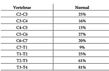
Vertebral level distribution of the osteoarthritis by articulation
Degrees of severity by articulation
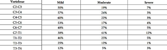
Source: “Characterization of bony changes localized to the cervical articular processes in a mixed population of horses”; by Kevin K. Haussler
The following statistics show a comparison between these two approaches.
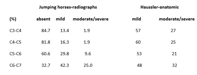
The only joint which shows a semblance of similarity is in C6-C7. This is all the more reason why the following quote on the limitations of radiographs is important:
“Radiographic anatomy of the articular process joints of the caudal cervical vertebrae in the horse on lateral and oblique projections”; by J M Withers 1, L C Voûte, G Hammond, C J Lischer 1Weipers Centre for Equine Welfare, University of Glasgow, Bearsden Road, Glasgow G61 1QH, UK. EQUINE VETERINARY JOURNAL Equine vet. J. (2009)
“Plain radiography is the standard imaging technique for investigation of diseases associated with the articular process joints (APJ) of the caudal neck; however, the radiographic anatomy of these structures on both lateral and oblique radiographic projections has not previously been described in detail.
Plain radiography is the primary imaging modality utilised in routine screening for osseous abnormalities of the cervical spine in the horse and a standard technique has been described previously (Whitwell and Dyson 1987; Butler et al. 2008). Routinely, this is performed with the horse standing and is limited to lateral-lateral projections (Whitwell and Dyson 1987; Butler et al. 2008).
Interpretation of cervical radiographs can be challenging due to the complex radiographic anatomy and overlap of structures on either side of the neck. Furthermore, because radiographs are 2D presentations of 3D structures, clinically significant lesions may be inadequately visualised when radiographs are obtained in one plane only. This limitation is illustrated in the difficulty in detecting axial enlargement of the articular processes on plain lateral-lateral radiographs (Butler et al. 2008)”.
In addition, there are numerous possible causes of a cervical problem, as well as the possibility that a horse may have several problems at the same time, as the next chart shows.
Cervical vertebra-number/type of lesions
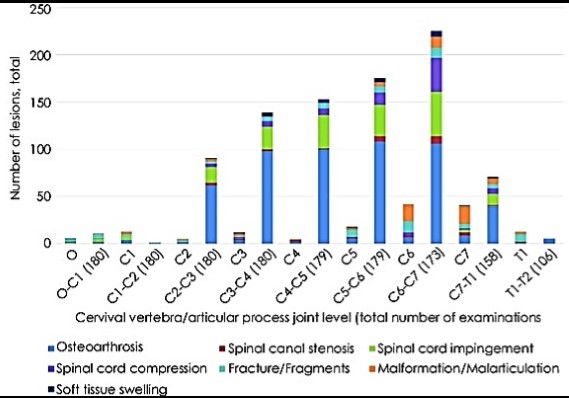
Source: “Computed tomography and myelography of the equine cervical spine: 180 cases (2013–2018)”, C. M. Lindgren, L. Wright, M. Kristoffersen, S. M. Puchalski; 2020, cc
Skeletal structures are also subject to changes in shape
There are also numerous types of osseous changes, as described next.
Prevalence of osseous changes
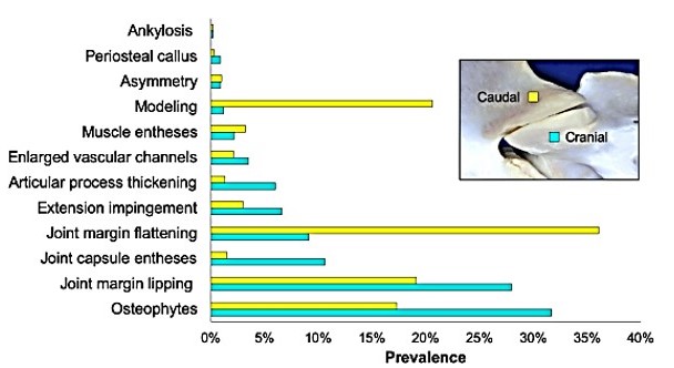
Note: Prevalence of the types of bony changes that are localized to the cranial (blue) and caudal (yellow) articular processes across all subjects.
Examples of osseous changes
Modelling
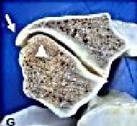
Flattening
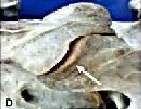
Lipping
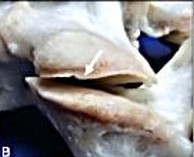
Osteophytes
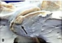
Source: “Characterization of bony changes localized to the cervical articular processes in a mixed population of horses”; by Kevin K. Haussler, cc
For the concerned horse owner, how is it possible to identify if this problem exists in their own horse(s)? What are the symptoms? The following quote elaborates.