Equi-note 17 Hyperflexion-rollkur-the horse is more conflictive because its airflow is blocked
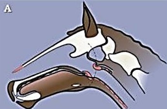
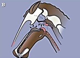
To understand the effects of rollkur it is necessary to view the effect on the horse’s windpipe.
Head and neck in moderate extension
(jowl angle 114°)
Head and neck in moderate flexion
(jowl angle 69°)
Notes:
1. Ascending arrows indicate direction of movement of soft palate; descending arrows indicate collapse of roof of the throat.
2. Jowl angle shown is 69°; when nose is vertical to ground jowl angle is 45°, but even this is far less than during rollkur.
Normal vs deformed windpipe/trachea
(Normal-nearly circular; deformed-elliptical and obstructed by intrusion of oesophagus)
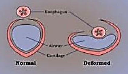
Source: “Why is ‘Rollkur’ Wrong? (part two) Continued observations on the Report of the FEI Veterinary and Dressage Committees’ Workshop on ‘The use of over-bending (“Rollkur”) in FEI Competition,” January 2006; wp
Equi-note 17
Hyperflexion-rollkur-the horse is more conflictive because its airflow is
blocked
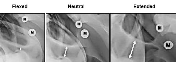
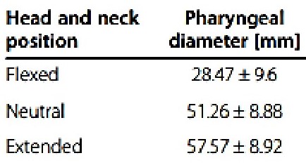
The following image and table shows what happens to the trachea when the horse’s head and neck are moved from flexion to neutral to extended.
“Objective classification of different head and neck positions and their influence on the radiographic pharyngeal diameter in sport horses”; by Li-mei Go1*† , Ann Kristin Barton1,2† and Bernhard Ohnesorge BMC Veterinary Research 2014, http://www.biomedcentral.com/1746-6148/10/118
Author’s note: “Lateral radiographs of the head taken in three head and neck positions/hnp. M are the metallic markers used to calculate magnification. Left image: flexed hnp; middle image: neutral hnp; right image: extended hnp.”
Pharyngeal diameter is length of white arrow
Source: “Objective classification of different head and neck positions and their influence on the radiographic pharyngeal diameter in sport horses” cc
Hyperflexion-rollkur-narrowing the trachea chokes the horse
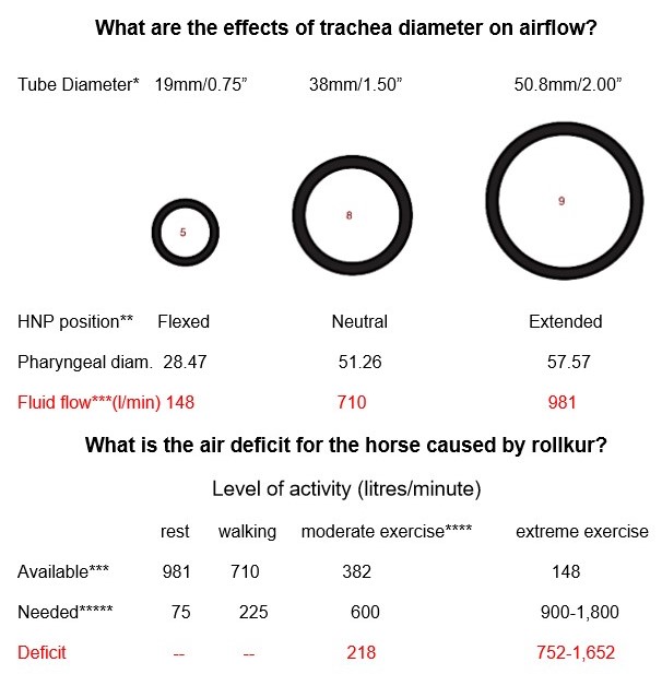
This is because a narrowed trachea has a significant effect by causing airflow reduction
The effect of changes in trachea diameter are shown next.
Deficit of 752-1,652 litres per minute = CHOKING
Notes:
* Kee Industrial products, Inc./cdn.simplifiedbulding.com.
** “ Why is ‘Rollkur’ Wrong? (part two) Continued observations on the Report of the FEI Veterinary and Dressage Committees’ Workshop on ‘The use of over-bending (“Rollkur”) in FEI Competition,” January 2006.
*** www.copely.com/tools/flow-rate-calculator/ Assumptions: PSI=1; pipe length=1m.
**** moderate exercise-assumes medium collection with trachea diameter of 40mm.
***** air available: 1)“Learn About Horse Lung Health” 2020, flairstrips.com; 2) Dr. J Cheetham VETMB, Dept. of Clinical Sciences; Cornell University College of Veterinary Medicine.
Hyperflexion-rollkur-narrowing the trachea blocks the airflow
How much air does the horse need?
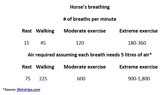
Further analysis of this vital issue is shown next.
“Airflow Mechanics, Upper Respiratory Diagnostics, and Performance-Limiting Pharyngeal Disorders”; John C. Janicek, DVM, MS, DACVSa Karissa M. Ketzner, DVM University of Missouri September 2008:
“Airway resistance (the pressure:flow ratio) is greatly affected by airway diameter
Cylindric tube resistance (R) can be calculated using the following formula (n = gas viscosity; L = length of trachea; r = radius of trachea):
R = (8 × nL) ÷ r 4
Gas viscosity, which affects the pressure:flow relationship, is constant. Because the radius is to the fourth power, a small difference in airway radius has a major impact on airway resistance. A 20% decrease in the airway radius doubles airflow resistance. In resting horses, two-thirds of the total airflow resistance during inspiration and expiration is in the upper airway.
During inspiration, negative pressure causes unsupported tissue to be drawn into the airway, resulting in upper airway narrowing; thus upper airway resistance during exercise becomes more than 80% of the total airflow resistance. During expiration while a horse exercises, upper airway resistance accounts for approximately 50% of the total airflow resistance. This is because the airway diameter is enlarged by positive pressure.
Most mammals can breathe by mouth during exercise. This provides a low-resistance pathway to accommodate the greater airflow requirement and thereby minimizing the work of breathing. However, the anatomic relationship of the larynx and caudal soft palate makes horses obligate nasal breathers. During exercise, the 20-fold increase in airflow while breathing must occur through the nose.
Other factors affecting upper airway function include head position and vascular engorgement
In resting horses, air entering the upper airway turns approximately 90 degrees to flow from the nasal passage into the trachea. During normal exercise, most horses straighten their head and neck. This increases the angle of the pharynx through which air flows and thereby reduces the work of breathing. Straightening of the airway allows a more direct route to the lungs. It also tends to stretch upper airway tissue, making it more resistant to collapse.
In some equestrian disciplines, horses carry their heads and necks in extended or flexed positions. For example, Thoroughbreds are raced with their head and neck extended. Whereas the head and neck are often flexed in Standardbreds and in sporting events such as show jumping and dressage.
Clinical signs of obstructive airway disease are often more evident when a horse’s head and neck are flexed. It has been demonstrated that when a horse’s head and neck are flexed, upper airway impedance is twice that of a horse exercising with its head extended. Head and neck flexion not only increases the work of breathing but also makes the upper airway tissue more prone to dynamic collapse”.
The next post analyzes the effect of increased speed on the trachea.